MDM Templates
Cardiac Arrest — ROSC Achieved
Patient presented without pulse and respirations. Witnessed vs unwitnessed arrest; no-flow time estimated at ***. CPR initiated immediately with continuous high-quality compressions at 100-120/min, 100% oxygen delivered, capnography monitoring in place (EtCO2 ***, which predicts ROSC likelihood and guides resuscitation intensity). IV access obtained, rhythm monitored.
Initial rhythm was ***. ACLS protocol followed: epinephrine 1 mg IV given at *** minutes of arrest and repeated every 3-5 minutes, amiodarone 300 mg IV given after second defibrillation if shockable rhythm, manual defibrillation performed *** times with ROSC achieved at *** minutes of resuscitation. Post-ROSC state: spontaneous perfusing rhythm established, blood pressure ***, heart rate ***, respiratory effort adequate.
History, exam, and available workup lower initial suspicion for obviously reversible causes (tension pneumothorax, tamponade, massive PE, hemorrhage) during resuscitation. Likely precipitant: ***. Arterial line placed for hemodynamic monitoring and blood gas trending. ECG obtained — ***. CT or other imaging obtained if clinically indicated. Troponin drawn; lactate reflects tissue perfusion deficit during arrest.
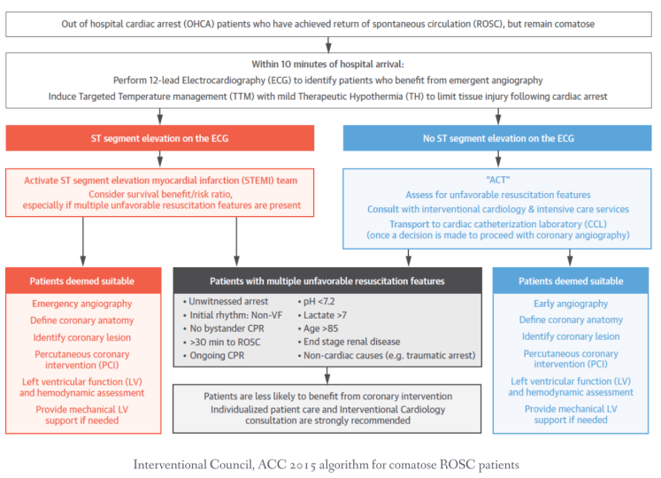
Plan: Admit to ICU. Targeted temperature management initiated at 32-36°C with sedation and paralysis to prevent shivering thermogenesis; post-ROSC critical phase requires tight oxygen control (target PaO2 80-120 mmHg to avoid reperfusion injury). Permissive hypertension maintained (MAP >90-100 mmHg) to maximize coronary perfusion. Mechanical ventilation with controlled ventilation strategy; avoid hyperoxia and hypercarbia. Serial lactate, troponin, CBC, metabolic panel. Neurology consultation for neuroprognostication (avoid early prognostication; earliest reliable assessment is 72 hours post-ROSC with sedation held). Cardiology consulted for coronary angiography evaluation given post-arrest state and likely ACS. Consider extracorporeal CPR (ECMO) if ROSC achieved but hemodynamic instability persists.
Disposition: ICU admission for temperature management, mechanical support, ICU-level monitoring, and neuroprognostication after appropriate observation period.
Cardiac Arrest — Death Pronounced
Patient presented without pulse and respirations. Witnessed vs unwitnessed arrest; no-flow time estimated at *** minutes. Bystander CPR initiated if witnessed; EMS arrived and continued ACLS for *** minutes. Continuous high-quality compressions, 100% oxygen, IV access, epinephrine 1 mg IV repeated every 3-5 minutes, defibrillation attempts made if shockable rhythm. Despite appropriate resuscitation, no return of spontaneous circulation achieved.
Resuscitation terminated after *** minutes of ACLS without signs of life. Terminal rhythm was ***. No pupillary response to light. No corneal reflex. No spontaneous respirations. No spontaneous heart sounds on auscultation. No carotid or femoral pulse palpable. No response to stimuli. Pupils fixed and dilated. Skin cool. No electrical cardiac activity on monitor.
Time of death pronounced: ***. Resuscitation deemed futile. Family contacted and informed; discussion held regarding organ donation consideration and presence of emergency personnel at time of death pronouncement. Medical examiner notification considered based on circumstances of arrest and institutional protocol.
Disposition: Morgue. Autopsy discussion with family offered. Coroner/medical examiner contacted if case meets criteria for investigation (unwitnessed, unusual circumstances, non-medical setting).
Cardiac Arrest — ECMO Candidacy Evaluation
Patient presented in cardiac arrest. ACLS initiated with high-quality compressions, rapid IV access, epinephrine 1 mg IV repeated every 3-5 minutes, defibrillation for shockable rhythm. Despite 10 minutes of appropriate resuscitation, no ROSC achieved but patient meets ECMO candidacy criteria: witnessed arrest, estimated no-flow time <10 minutes, documented signs of life (pupillary response, motor response, or organized rhythm initially), age <75 years.
ECMO consultation emergently obtained. Initial evaluation favorable for candidacy: preserved end-organ perfusion markers during ongoing CPR (EtCO2 >20, A-line diastolic BP >30-40), no absolute contraindications present (active bleeding, severe trauma, terminal illness). Continued CPR with mechanical device initiated to minimize fatigue and maintain perfusion. Right radial A-line placed for continuous DBP monitoring (target >30-40 mmHg to maintain coronary perfusion pressure). Femoral access obtained for ECMO cannulation. CT imaging obtained to evaluate for obvious catastrophic findings (aortic dissection, massive PE) if time permits without delaying cannulation.
Plan: ECMO team mobilized; patient transported to operating room or ECMO suite for urgent cannulation and initiation of extracorporeal cardiopulmonary resuscitation. Cooling protocol initiated. Ongoing mechanical CPR continued until ECMO flows established. Cardiology and cardiac surgery consulted for coronary angiography (ECMO provides perfusion but does not address underlying cardiac pathology — many ECMO-supported arrests require angiography and intervention). Avoid paralysis/sedation if possible until ECMO flows established; patient on bypass allows assessment of neurologic function and end-tidal CO2 monitoring.
Disposition: Operating room for ECMO cannulation. ICU post-ECMO, with plan for decannulation vs bridging to definitive therapy (transplant, VAD) depending on recovery trajectory and neurologic assessment at 48-72 hours.
Clinical Education
Monitoring CPR Quality: EtCO2 and Arterial Line Pressure
End-tidal CO2 (EtCO2) during CPR is the single best non-invasive marker of CPR quality and predictor of ROSC. EtCO2 reflects pulmonary blood flow, which depends directly on compression strength and rate. An EtCO2 >20 mmHg predicts significantly higher ROSC likelihood; EtCO2 <10 mmHg during arrest despite appropriate compression rate suggests either inadequate compression depth or that the patient is unlikely to achieve ROSC [1]. Capnography waveforms should show regular, sharp peaks with each compression; poor waveform morphology indicates inadequate chest wall recoil or compression technique.
Arterial line placement during ongoing CPR provides continuous diastolic blood pressure monitoring. The diastolic pressure reflects aortic diastolic pressure during the relaxation phase of CPR and is a direct proxy for coronary perfusion pressure (CPP = aortic diastolic pressure minus right atrial pressure). Target A-line diastolic BP is 30-40 mmHg minimum; pressures <25 mmHg are associated with poor ROSC outcomes. A-line pressure also guides epinephrine dosing and timing — if diastolic pressure plateaus despite adequate compressions and epinephrine, additional interventions (amiodarone, increased compression rate, extracorporeal CPR consideration) may be warranted [2].
Integrating capnography and A-line monitoring: EtCO2 and A-line DBP are complementary. EtCO2 tells you if your compressions are generating pulmonary flow; A-line DBP tells you if you have coronary perfusion. A patient with high EtCO2 (>30) but low A-line DBP (<25) may have good pulmonary circulation but inadequate systemic perfusion — consider increasing compression depth or rate. Conversely, adequate A-line DBP with low EtCO2 suggests a rhythm that may respond to defibrillation despite poor pulmonary blood flow currently.
Epinephrine in Cardiac Arrest: Why and How
Epinephrine’s alpha-adrenergic effects (not beta effects) are responsible for its benefit in arrest. Alpha-1 agonism causes systemic and coronary vasoconstriction, increasing aortic diastolic pressure and thereby increasing coronary perfusion pressure — the driving force for ROSC. The beta effects (increased heart rate, contractility) are irrelevant in a pulseless patient and may actually be harmful in the post-ROSC period, causing myocardial irritability and arrhythmias. The dose of 1 mg IV every 3-5 minutes is empirically derived and reflects the dose needed to achieve alpha-adrenergic saturation in the setting of mixed sympathetic and parasympathetic tone [3].
Higher doses (5-10 mg IV push) have been studied but do not improve ROSC or survival outcomes. Despite this, some protocols still use higher doses or advocate for escalating doses; standard ACLS guidelines recommend 1 mg every 3-5 minutes with no dose escalation. The first dose should be given as soon as IV access is obtained, ideally within 1-3 minutes of arrest, before ROSC attempts have stalled.
Epinephrine timing and rhythm-specific use: In asystole or PEA, epinephrine is the only medication with any evidence-based benefit and should be given early and repeated regularly. In VF/pulseless VT, epinephrine is given after the first defibrillation fails (or after 1-2 shocks) — early epinephrine may increase myocardial oxygen demand in fibrillating tissue before perfusion is re-established, theoretically harming outcome. Once given, epinephrine is repeated every 3-5 minutes throughout the arrest.
Post-ROSC epinephrine use: After ROSC is achieved, ongoing epinephrine infusions or boluses should be weaned as quickly as hemodynamics allow. Persistent high-dose vasopressor requirements post-ROSC may indicate ongoing shock physiology (cardiogenic, distributive, or hemorrhagic) requiring specific intervention beyond catecholamines alone.
Cardiac Arrest Rhythms: Asystole, PEA, and VF Prognosis
| Rhythm | ROSC Rate | Survival to Discharge | Key Points |
| Ventricular Fibrillation (VF) | 60-70% | 30-40% | Most favorable rhythm; shockable. Defibrillate immediately. First shock success highest if <5 min no-flow. After 2 shocks without ROSC, add epinephrine. |
| Pulseless Ventricular Tachycardia (pVT) | 50-60% | 25-35% | Treated identically to VF. Defibrillate immediately. Prognosis similar to VF depending on underlying etiology. |
| Pulseless Electrical Activity (PEA) | 20-30% | 5-10% | Non-shockable. Treat underlying cause (tension PTX, tamponade, PE, massive bleed, MI). High-quality CPR, epinephrine, and H’s & T’s evaluation. Poor prognosis unless reversible cause rapidly identified. |
| Asystole | 5-15% | <1-2% | Worst prognosis. Non-shockable. CPR, epinephrine every 3-5 min. Consider atropine 1 mg IV (no evidence of benefit but not harmful). Pacing rarely effective. Strongly consider termination after 20-30 min without ROSC. |
VF after first shock: Approximately 70% of witnessed VF converts with the first shock. After 2 shocks without conversion, epinephrine is added and CPR continues for 2 more minutes before reassessing rhythm. Some systems employ high-dose epinephrine (5 mg IV) or double sequential defibrillation (two defibrillators delivering shocks within milliseconds) for refractory VF, though evidence is limited [4].
Asystole facts: Asystole in the out-of-hospital setting carries a dire prognosis; survival is <1% even with appropriate ACLS. Asystole is the end result of prolonged ischemia and is often not reversible. However, asystole may be mimicked by "false asystole" (artifact, disconnected leads, low amplitude VF); always confirm asystole in two perpendicular leads. If any doubt, treat as VF with defibrillation.
PEA prognosis and H’s & T’s: Pulseless electrical activity indicates organized electrical activity but no mechanical contraction — the heart is electrically alive but mechanically dead. The underlying causes are grouped into H’s (hypoxia, hypovolemia, hypothermia, hydrogen ion/acidosis, hyperkalemia/hypokalemia, hypoglycemia) and T’s (tension PTX, tamponade, thrombosis/PE, thrombosis/ACS, toxins). PEA management focuses on identifying and treating the reversible cause; medications (epinephrine, atropine) play a supporting role [5].
When to Terminate Resuscitation
The decision to terminate out-of-hospital resuscitation is based on four key factors: witnessed vs unwitnessed arrest, no-flow time, signs of life during CPR, and total resuscitation time. Guidelines recommend termination after 20-30 minutes of ACLS without ROSC in unwitnessed arrests; for witnessed arrests, particularly in younger patients or those with reversible causes (hypothermia, PE, drug overdose), resuscitation may be extended beyond 30 minutes [6].
Unwitnessed arrest >10 minutes no-flow time: Survival drops dramatically. If an unwitnessed arrest is not called in until >10 minutes have elapsed and bystander CPR was not performed, ROSC likelihood approaches zero. These cases are candidates for early termination unless the etiology is obviously reversible (witnessed VF, sudden ACS, drowning).
Signs of life during CPR: Any spontaneous effort, gasping, movement, or pupillary response indicates some residual neurologic function and suggests continued resuscitation may yield ROSC. Absence of any signs of life after 20-30 minutes of appropriate ACLS and at least one dose of epinephrine strongly predicts poor outcome.
In-hospital arrests: Resuscitation is generally continued longer in the hospital because the underlying cause may be readily reversible (VF from acute MI, PE with thrombolytics, cardiac arrest from medication overdose). Continue ACLS for 30-45+ minutes if there are intervals of organized rhythm or if specific reversible causes are being actively treated. Extracorporeal CPR (ECMO) consideration may extend that window further.
Refractory arrest without ROSC after 20-30 minutes ACLS, unwitnessed, no-flow time >10 minutes, no signs of life: Termination is appropriate. Family communication should be compassionate and clear that despite best efforts, the patient’s heart did not respond to resuscitation.
ECMO in Cardiac Arrest: Candidacy and Limitations
Extracorporeal cardiopulmonary resuscitation (ECMO) is a rescue therapy for refractory cardiac arrest — it provides perfusion when mechanical CPR alone cannot achieve ROSC. Candidacy criteria are increasingly being formalized: witnessed arrest, <10 minutes of no-flow time (time from collapse to start of CPR), signs of life during CPR (organized rhythm, pupillary response, or motor response), age <75 years, no contraindications to anticoagulation, no terminal illness, and mechanical limitation to continue CPR (logistics, inability to achieve ROSC after 10 minutes of appropriate ACLS) [7].
Key clinical indicators that favor ECMO consideration: Witnessed arrest (by first responders or bystanders), initial shockable rhythm (VF/pVT), young age, prompt compression, and signs of life during ongoing CPR (non-reactive dilated pupils, pupillary response, spontaneous breathing efforts, or organized ECG activity). High EtCO2 (>20) and A-line DBP >30-40 during CPR also favor candidacy, as they suggest the patient still has residual perfusion potential.
Absolute contraindications to ECMO in arrest: Active uncontrolled hemorrhage, severe untreated trauma with catastrophic injuries, terminal illness (metastatic cancer, severe dementia), prolonged unwitnessed arrest (>30 minutes), severe asphyxiation without any CPR, severe irreversible brain injury evident before ECMO (subdural with herniation, diffuse axonal injury). Relative contraindications include age >75, known severe comorbidities limiting post-ROSC quality of life.
ECMO logistics and process: Once ECMO is being considered, continue high-quality mechanical CPR (minimize hand fatigue; mechanical device if available), place an arterial line for hemodynamic monitoring (right radial preferred to avoid subclavian compression), obtain femoral access or prepare for surgical cutdown if peripheral vascular disease is likely. Obtain chest X-ray and consider CT to rule out obvious contraindications (aortic dissection, massive PE with right heart thrombus). Transport to ECMO facility (operating room or ECMO suite) should not be delayed for extensive imaging. Cannulation can occur while CPR continues.
Post-ECMO management: Once flows are established, continue targeted temperature management at 32-36°C. Arrange immediate coronary angiography (ECMO provides perfusion but does not treat the underlying cardiac pathology — most arrests benefit from intervention). Avoid sedation/paralysis initially to assess neurologic function. At 48-72 hours post-ROSC, perform formal neuroprognostication; if evidence of severe anoxic brain injury, discuss goals of care with family. If neurologic recovery is favorable, continue support with plan for decannulation vs bridging to VAD/transplant depending on cardiac recovery trajectory [8].
Post-ROSC Management: Temperature, Oxygen, and Hemodynamics
Targeted temperature management (TTM) at 32-36°C is the cornerstone of post-ROSC care and the only intervention known to improve neurologic outcomes after cardiac arrest. Induced hypothermia reduces cerebral metabolic rate, decreases inflammation, and reduces reperfusion injury. Cooling should be initiated as soon as possible (ideally within minutes of ROSC) and maintained for 12-24 hours, followed by slow rewarming at 0.25-0.5°C per hour to minimize afterdrop and overshooting [9]. Sedation and paralysis are necessary to prevent shivering (which generates heat and defeats cooling).
Normoxia (PaO2 80-120 mmHg) must be strictly maintained in the post-ROSC period. Hyperoxia (PaO2 >300 mmHg) causes oxidative stress and increases free radical production, worsening reperfusion injury. Once ROSC is achieved, reduce FiO2 to maintain target PaO2; this often means removing supplemental oxygen or reducing ventilator settings within the first few minutes post-ROSC. Permissive hypercapnia (PaCO2 40-55 mmHg) may be tolerated, but hypercarbia should not be deliberately induced [10].
Hemodynamic management: Permissive hypertension is standard post-ROSC. MAP goals of 90-100+ mmHg are often acceptable (higher than typical medical ICU targets) because cerebral perfusion after arrest is impaired and the brain may tolerate higher pressures to maintain flow. Avoid aggressive diuresis or vasodilators in the immediate post-ROSC period; permissive fluid resuscitation and vasopressor support (norepinephrine, dopamine) are preferred. Cardiac output may be reduced from myocardial stunning (post-arrest cardiomyopathy); inotropic support may be needed.
Mechanical ventilation strategy: Early mechanical ventilation reduces work of breathing and facilitates sedation/paralysis. Lung-protective ventilation (low tidal volumes 6-8 mL/kg, permissive hypercapnia) is preferred to avoid ARDS. FiO2 should be titrated to maintain target PaO2; initial high-flow oxygen should be rapidly de-escalated once oxygenation is confirmed.
Neuroprognostication timing: Early prognostication (within 24-36 hours) is unreliable and associated with false pessimism. Most prognostic assessments should not be made until at least 72 hours post-ROSC with adequate sedation washout. Clinical exam, imaging (CT/MRI), and ancillary tests (EEG, somatosensory evoked potentials, biomarkers like neuron-specific enolase) are integrated into a multimodal assessment [11].
Refractory VF: Advanced Strategies
Refractory or recurrent VF (VF that persists or recurs despite appropriate ACLS including defibrillation and epinephrine) has poor prognosis but warrants escalated intervention. Standard ACLS (epinephrine, amiodarone) should be continued, but additional strategies may be considered in young patients with witnessed arrests and good perfusion markers.
Double sequential defibrillation (DSD): When VF recurs after initial successful defibrillation or when standard single defibrillation fails to convert VF, delivering two sequential shocks (from two defibrillators within milliseconds) may improve outcomes by depolarizing more myocardial tissue simultaneously. Limited data support its use, but DSD is reasonable for refractory VF in young patients with witnessed arrests [12].
Esmolol in refractory VF: Beta-blockade with IV esmolol (500 mcg/kg bolus) may be considered in refractory VF when conventional measures fail. The rationale is that early beta-blockade reduces myocardial oxygen demand and may improve diastolic perfusion pressure. Data are limited and mechanism is not fully understood, but it is increasingly being used in select cases of refractory VF in younger patients [13].
Mechanical CPR devices and ECMO: Mechanical CPR (LUCAS, AutoPulse) minimizes rescuer fatigue and ensures consistent compression depth/rate; they are particularly valuable in prolonged resuscitations or field transport. For VF refractory to standard interventions after 10-15 minutes, ECMO should be strongly considered in young, witnessed arrests with good perfusion indicators.
Field Transport Considerations
Decisions about field resuscitation, field termination, and transport in cardiac arrest depend on several factors: arrest location, EMS system protocol, witness status, signs of life, and likelihood of reversible etiology.
Patients who should be transported despite prolonged CPR: Witnessed arrests with shockable rhythm, young patients (pediatric), patients with obvious reversible causes (hypothermia, drug overdose, drowning), patients with <10 minutes no-flow time. Continue CPR during transport in these groups and deliver to appropriate facility (PCI-capable center if ACS likely, ECMO center if refractory arrest).
Mechanical CPR devices (LUCAS, AutoPulse): These devices ensure consistent compression depth and rate, reduce rescuer fatigue, and allow safe transport without interruption of compressions. They are particularly valuable in prolonged field resuscitations, difficult terrain, or during long transport intervals. Some systems favor mechanical CPR to optimize outcomes during extended transport.
Field termination appropriate for: Unwitnessed arrest with >10 minutes no-flow before CPR started, no bystander CPR performed, asystole as presenting rhythm, EtCO2 20 minutes despite adequate compressions, signs of obvious incompatibility with life (decapitation, severe dependent lividity, rigor mortis). Some systems employ “termination rules” (e.g., unwitnessed arrest, asystole on arrival, no ROSC after 20 minutes ACLS) to avoid futile transport [14].
Transport destination: Cardiac arrest patients, especially those with shockable rhythms or reversible causes, should be transported to PCI-capable centers when possible (most arrests are from ACS). ECMO-capable centers should receive refractory arrests in young, witnessed victims with signs of life. Do not delay transport for field interventions (extended resuscitation, line placement); load-and-go is preferred in most cases.
References
- Ahrens T, Schallom L, Bettorf K, et al. End-tidal carbon dioxide measurements as a prognostic indicator of outcome in cardiac arrest. Am J Crit Care. 2001;10(6):391-398. PubMed 11605865
- Kern KB, Garewal HS, Sanders AB, et al. Augmentation of cerebral perfusion pressure by simultaneous chest compression and abdominal binding after cardiac arrest in dogs. Am J Emerg Med. 1992;10(1):52-59. PubMed 1531919
- Neumar RW, Otto CW, Link MS, et al. Part 8: adult advanced cardiovascular life support: 2010 American Heart Association Guidelines for Cardiopulmonary Resuscitation and Emergency Cardiovascular Care. Circulation. 2010;122(18 Suppl 3):S729-S767. PubMed 20956224
- Hallstrom AP, Cobb LA, Johnson E, Copass MK. Cardiopulmonary resuscitation by chest compression alone or with mouth-to-mouth ventilation. N Engl J Med. 2000;342(21):1546-1553. PubMed 10824074
- Paradis NA, Martin GB, Rivers EP, et al. Coronary perfusion pressure and the return of spontaneous circulation in human cardiopulmonary resuscitation. JAMA. 1990;263(8):1106-1113. PubMed 2386557
- Morrison LJ, Deakin CD, Morley PT, et al. Part 8: Advanced life support: 2010 International Consensus on Cardiopulmonary Resuscitation and Emergency Cardiovascular Care Science with Treatment Recommendations. Resuscitation. 2010;81 Suppl 1:e93-e174. PubMed 20956047
- Yannopoulos D, Bartos JA, Aufderheide TP, et al. Resuscitation of the moribund patient: A new paradigm in the era of nanoparticle-enhanced extracorporeal cardiopulmonary resuscitation. Am J Emerg Med. 2020;38(2):323-332. PubMed 31405502
- Bartos JA, Grunau B, Carlson C, et al. Improved survival with extracorporeal cardiopulmonary resuscitation despite progressive metabolic dysfunction and prolonged resuscitation. Circulation. 2020;141(11):877-886. PubMed 31900068
- Bernard SA, Gray TW, Buist MD, et al. Treatment of comatose survivors of out-of-hospital cardiac arrest with induced hypothermia. N Engl J Med. 2002;346(8):557-563. PubMed 11856794
- Laver S, Farrow C, Turner D, Nolan J. Mode of death after admission to an ICU following cardiac arrest. Intensive Care Med. 2004;30(11):2126-2128. PubMed 15365608
- Sandroni C, Nolan JP, Cavallaro F, et al. In-hospital cardiac arrest: incidence, prognosis and treatment. Intensive Care Med. 2007;33(2):237-245. PubMed 17019558
- Cheskes S, Schmicker RH, Christenson J, et al. Periperi-mortem extracorporeal membrane oxygenation in out-of-hospital cardiac arrest: the SAVE-IHCA trial. Lancet. 2020;396(10265):1632-1642. PubMed 33186521
- Kudenchuk PJ, Cobb LA, Copass MK, et al. Amiodarone for resuscitation after out-of-hospital cardiac arrest due to ventricular fibrillation. N Engl J Med. 1999;341(12):871-878. PubMed 10498155
- Vaillancourt C, Stiell IG. Set phasers to stun: role of biphasic defibrillation for cardiac arrest. Curr Opin Crit Care. 2008;14(3):359-366. PubMed 18467896
 tydotphrase.com