Endotracheal intubation
The patient required endotracheal intubation.
The patient was given
Etomidate approx. 0.3 mg per kilogram
Rocuronium approx. 1 mg per kilogram
Once the patient was adequately sedated and paralyzed, a Mac 4 laryngoscope was used to directly visualize the cords.
Using this direct visualization, a 7.5 endotracheal tube was then passed easily through the cords.
This tube was inserted to 21 cm at the lip.
There was excellent color change on the end-tidal CO2 monitor. The patient was easily and adequately ventilated. There were excellent breath sounds bilaterally with no breath sounds heard over the epigastrium.
The tube was secured in the standard fashion. The patient tolerated this procedure well and there were no complications.
Post-intubation chest x-ray demonstrates excellent endotracheal tube placement.
PEARLS
- Airway Algorithm
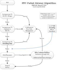
- (SG) Standard Geometry, (HA) HyperAngulated blades
- Weingart argues you should bougie immediately so you can become a master of one technique, I think two.
- Techniques
- Pre-intubation Medications
- RSI
-
Most Common
-
Etomidate 20mg (0.30 mg/kg) + rocuronium 80mg (1 mg/kg)
-
-
3:2:1 regimen
-
Fentanyl (3 mcg/kg) + ketamine (2 mg/kg) + rocuronium (1 mg/kg). Given in rapid succession in that order.
-
Source: Lyon R. et al. Critical Care 2015
-
-
-
- Hemodynamic compromise
- 1:1:1 regimen
- fentanyl (1 mcg/kg IV), ketamine (1 mg/kg IV), rocuronium (1 mg/kg) in that order
- consider Half Dose etomidate (0.15 mg/kg) instead of ketamine
- Consider no paralytic
- fentanyl (1 mcg/kg IV), ketamine (1 mg/kg IV), rocuronium (1 mg/kg) in that order
- 1:1:1 regimen
- Delayed Sequence Intubation
- Indication: AMS and thrashing or otherwise unable to preoxygenate (pretty much Procedural Sedation)
- 1. Ketamine 1-2mg/kg
2. NIPPV or bag-valve mask with PEEP valve at 10 cmH20 until SpO2 is <95%
3. Paralyze with Rocuronium (1 mg/kg) while providing apneic oxygenation with 15 L/min O2 via nasal prong
4. Intubate
- Bicarb (in patients known pH <7.15)
- If patient is known to be extremely acidodic, transient apnea will only make them more acidodic and their pressors may stop working
- RSI
- Post Intubation
- Sedation
- Fentanyl 1mcg/kg IV bolus followed by 0.5-1.0 mcg/kg/hr
- Generally 50-100mcg safe for adult
- Versed 0.05 mg/kg bolus followed by 0.025 mg/kg/hr
- Generally 2-5mg bolus followed by 1-3mg/hr safe for adult
- Propofol 0.5mg/kg bolus followed by 10-50mcg/kg/min
- Generally (100kg adult)
- Unstable (Trauma, ROSC, Septic Shock) start at 10mcg/kg/min = 1mg/min = 60mg/hr
- Stable: Double the unstable
- Can bolus 20-40mg like in a procedural sedation
- Generally (100kg adult)
- Fentanyl 1mcg/kg IV bolus followed by 0.5-1.0 mcg/kg/hr
- Ventilator Settings
- Generally (75kg)
- TV 400-500ml (6-8ml/kg IBW), RR 12, PEEP 5 (titrate up to 10), FiO2 start at 100% (wean to 40-60% with goal SpO2 > 94%), I:E 1:2
- Generally (75kg)
- Sedation
- Extubation
- Checklist
- Resolution of process necessitating intubation
- Respiratory:
- RSBI < 80
RR < 35
No increased WOB - FiO2 < 40% with O2 saturation > 94%
PEEP 5 or less
- RSBI < 80
- Patient hemodynamically stable without pressor requirement
- Awake, Alert, Following Commands, and without recent seizure acitvity
- How to
- Position the patient sitting upright (or on side)
- Prepare a high-flow oxygen mask and have a full airway cart nearby
- Remove secretions from oropharynx with Yankauer suction
- Remove ETT securing straps
- At end inspiration, deflate the cuff and quickly and smoothly remove the ETT during exhalation
- Place on high-flow oxygen mask and monitor the patient closely for 30 minutes minimum
- https://emcrit.org/wp-content/uploads/2010/11/SaraGray-ED-Extubation.pdf
- Extubation Note: Patient had resolution of process necessitating intubation with low suspicion for inability to maintain airway henceforth. They were able to maintain oxygenation and ventilation on low ventilator settings. Patient was positioned upright, secretions were removed from oropharynx, ETT was deep suctioned, at end inspiration cuff was deflated and tube was removed during exhalation. Patient placed on high flow O2 and monitored for 30+ minutes. Remained stable.
- Checklist
- Endotracheal Tubes
- Tube depth: Often taught to be best at 3x the tube size. 21 (shorter girl) -24 (tall man) from central incisors. Stone et al 2005. Airway Management
- Tube Sizing
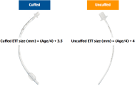
- Only ever use uncuffed in neonate…
- Laryngoscopy Blades
- Video Advantages
- Teaching obviously
- Can jump straight from video to DL in a SA blade if camera is obstructed by saliva etc
- Obvious increased ability to see anterior airways
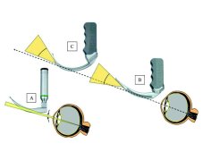
- Mac 3 advantages:
- fulcrum of torque being closer allows you to resist rotational force patient head on blade and lift easier
- Video Advantages
-
Blind nasotracheal intubation:
-
Consider when obstruction present: severe angioedema, fracture or recent surgery.
-
Consider when patient cannot tolerate laying back: Severely dyspneic patients with CHF, COPD, or asthma who are awake often cannot remain supine but may tolerate nasotracheal intubation in the sitting position.
- 1-2 cc of the lidocaine in each nare
Lubricate and place a NPA
Insert the ET tube you orient the bevel of the tube towards the septum (to avoid injury to the inferior turbinate)(6-7 cm feel a “give”)
Note fogging as just above cords and advance upon inspiration
Upon passing through the vocal cords, stridulous breathing and reflex coughing can be noted
Nare to the tip of the tube ~28 cm in males and 26 cm in females- Consider pretreatment with phenylephrine drops or oxymetazoline spray
-
 tydotphrase.com