Cardiopulmonary ECHO
Due to this patient’s [Shortness of Breath/Chest Pain], a focused bedside sonography for pulmonary and cardiac function was indicated.
Bedside ultrasonography was utilized to complete this procedure.
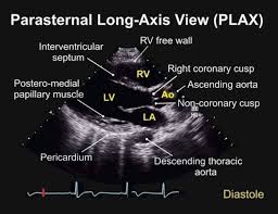
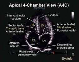
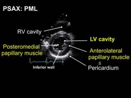
Cardiac evaluation: Both the parasternal and subxiphoid approaches were used to visualize the heart and pericardial sac. There is no evidence of gross wall motion abnormality. There is no evidence of pericardial effusion.
Pulmonary evaluation: No significant pleural effusions and < 3 B-lines per Intercostal Space on anterior exam
Images were obtained and filed for further review if necessary.
The patient tolerated this procedure well and there were no complications.
Trauma FAST
Due to this patient’s trauma, a focused abdominal sonography for trauma was indicated. Bedside ultrasonography was utilized to complete this procedure.
Abdominal evaluation:
Morison’s pouch: There is no evidence of free fluid in Morrison pouch.
Splenorenal junction: There is no free fluid visible.
Bladder: Views of the bladder were obtained in both transverse in sagittal planes. There was no e/o extravasation or surrounding free fluid
Limited echocardiogram: Both the parasternal and subxiphoid approaches were used to visualize the heart and pericardial sac. There is no evidence of gross wall motion abnormality. There is no evidence of pericardial effusion.
Images were obtained and filed for further review if necessary.
The patient tolerated this procedure well and there were no complications.
AAA r/o
Limited bedside ultrasound done by ED MD
Indication: Dizziness and hypotension
Type of Scan: Transabdominal
Areas Examined: Aorta from SMA to Iliac bifurcation
Findings: Aorta appeared less than 3 cm from SMA to iliac bifurcation in both transverse and longitudinal views, iliacs both appeared less than 1.5 cm in transverse view
Impression: No sonographic evidence of aortic or iliac dissection
- General Tips
- Aorta >3cm = bad, Iliacs >1.5cm = bad
- Most are infrarenal
- Measure outer wall in case of thrombus within aneurysm (don’t mess-up and stop at the thrombus)
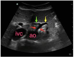
- Seagull sign of Splenic (yellow) and hepatic (green) vein branch-point from Celiac trunk (red)
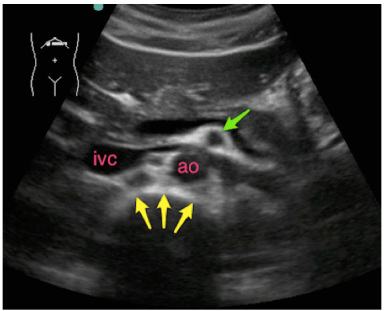
- Clock-Mantle sign of SMA (green)
Retinal Detachment r/o
Retinal Detachment r/o
Limited bedside ultrasound done by ED
Indication: Eye Pain
Type of Scan: Ocular
Areas Examined: RIGHT eye
Findings: Retinal contour NORMAL
Impression: NO sonographic evidence of retinal detachment
 tydotphrase.com