Last reviewed: March 2026
Contents
MDM Templates
Rash — Benign Viral Exanthem
Child presents with diffuse maculopapular rash in the setting of viral symptoms. Well appearing, no toxic appearance, no mucosal involvement, no petechiae or purpura. Rash blanches with pressure. No joint swelling, no facial or lip edema.
Presentation consistent with benign viral exanthem. Not consistent with Kawasaki disease (lacks sufficient diagnostic criteria), meningococcemia (no petechiae, well appearing), Stevens-Johnson syndrome (no mucosal involvement or target lesions), or ITP (no petechiae/purpura).[1]
Plan: Supportive care with antipyretics and antihistamines for pruritus. Discharge with PCP follow-up. Return for new petechiae or purpura, mucosal involvement, persistent fever beyond 5 days, joint swelling, or toxic appearance.
Rash — Kawasaki Disease Concern
Child presents with fever for *** days and clinical features concerning for Kawasaki disease. Findings include polymorphous rash, conjunctival injection, oral mucosal changes, extremity changes, and/or cervical lymphadenopathy.
Presentation meets criteria for classic Kawasaki disease (fever ≥5 days plus ≥4 of 5 principal criteria) or raises concern for incomplete Kawasaki (fever ≥5 days with 2-3 criteria and laboratory support). Untreated Kawasaki carries 25% risk of coronary artery aneurysm. Not consistent with benign viral exanthem, scarlet fever, or drug reaction given the clinical constellation.[2]
Plan: Echocardiogram. IVIG 2 g/kg plus high-dose aspirin initiated per guidelines. Pediatric cardiology consulted. Admit for treatment and monitoring.
If incomplete Kawasaki suspected (fever ≥5 days with 2-3 criteria): CRP ≥3 mg/dL or ESR ≥40 with ≥3 supplementary lab criteria (anemia, platelets >450k after day 7, albumin ≤3, elevated ALT, WBC ≥15k, urine WBC ≥10/hpf) supports the diagnosis and treatment.[2]
Rash — Urticaria / Allergic Reaction
Child presents with urticarial rash — raised, erythematous, pruritic wheals that blanch with pressure. No angioedema, no respiratory distress, no hypotension, no mucosal swelling. Individual lesions migratory and transient.
Presentation consistent with acute urticaria without anaphylaxis. No airway compromise, no cardiovascular instability. Not consistent with urticarial vasculitis (lesions not fixed or painful), serum sickness (no fever, arthralgias), or anaphylaxis.[1]
Plan: Cetirizine or diphenhydramine for symptom control. Discharge with PCP follow-up. Return for lip or tongue swelling, difficulty breathing, throat tightness, vomiting, or recurrence with identified trigger.
If anaphylaxis (urticaria plus respiratory distress, hypotension, or GI symptoms): IM epinephrine 0.01 mg/kg (max 0.3 mg pediatric). Observe minimum 4 hours post-treatment. Discharge with EpiPen prescription and allergy referral.
Rash — Petechial / Purpuric
Child presents with petechial or purpuric rash — non-blanching lesions. This mandates evaluation for serious underlying pathology regardless of clinical appearance.
Differential includes meningococcemia (rapidly progressive, toxic, febrile), ITP (well child with isolated thrombocytopenia), HSP (palpable purpura on lower extremities with joint and abdominal pain), leukemia (constitutional symptoms, cytopenias, hepatosplenomegaly), and DIC. Petechiae isolated above the nipple line from coughing or vomiting are a benign mechanical cause but require clinical judgment.[1]
Plan: CBC with differential and peripheral smear, coagulation studies, blood culture. If febrile with petechiae and any toxic appearance, empiric ceftriaxone immediately before results return. Disposition based on underlying etiology.
Clinical Education
Approach to Pediatric Rash
The critical first question: does this rash blanch? Non-blanching rashes (petechiae, purpura) require urgent evaluation for meningococcemia, ITP, HSP, and leukemia. Blanching rashes are far more commonly benign but still require systematic assessment.[1]
Pattern recognition drives the differential. Maculopapular and diffuse: viral exanthem, drug reaction, Kawasaki. Vesicular: varicella, HSV, hand-foot-mouth. Urticarial: allergic reaction, serum sickness. Petechial/purpuric: meningococcemia, ITP, HSP, leukemia. Target lesions: erythema multiforme, SJS.
Red flags in any pediatric rash: toxic appearance, petechiae with fever, mucosal involvement (SJS/TEN), rapidly spreading purpura, desquamation with fever (Kawasaki, TSS), and blistering >10% BSA.
Kawasaki Disease
Kawasaki disease is the leading cause of acquired heart disease in children in the developed world. Untreated, 25% develop coronary artery aneurysms. With timely IVIG within 10 days of fever onset, this drops to <5%. The diagnosis is entirely clinical — there is no confirmatory test.[2]
Classic criteria: Fever ≥5 days PLUS ≥4 of 5 principal features — (1) bilateral non-exudative conjunctival injection, (2) oral mucosal changes (strawberry tongue, cracked/erythematous lips), (3) polymorphous rash, (4) extremity changes (edema, erythema, later desquamation), (5) cervical lymphadenopathy (≥1.5 cm, usually unilateral).
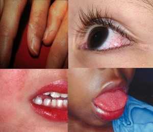
Incomplete Kawasaki is the diagnostic trap. Children <1 year are most likely to present incompletely and most likely to develop coronary aneurysms. Fever ≥5 days with only 2-3 criteria plus supportive labs (CRP ≥3 or ESR ≥40 with ≥3 supplementary criteria) warrants treatment. When in doubt, get an echo and treat.[2]
Treatment: IVIG 2 g/kg single infusion plus high-dose aspirin (80-100 mg/kg/day divided QID) until afebrile, then low-dose aspirin (3-5 mg/kg/day) for 6-8 weeks. Retreatment with IVIG if fever persists >36 hours after initial dose. Echocardiogram at diagnosis, 2 weeks, and 6-8 weeks.
Hand-Foot-Mouth Disease
HFMD is a clinical diagnosis. Caused by Coxsackievirus A16 and Enterovirus 71. Classic triad: oral vesicles/ulcers (painful, on tongue and buccal mucosa), vesicular lesions on hands and feet (palms and soles), and low-grade fever. Coxsackievirus A6 causes atypical widespread distribution.[3]
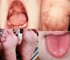
Herpangina is a related enteroviral syndrome — posterior oropharyngeal vesicles/ulcers with fever and odynophagia, WITHOUT hand and foot lesions. Higher fevers than HFMD. Both are self-limited (7-10 days). Key management concern is dehydration from painful oral intake.
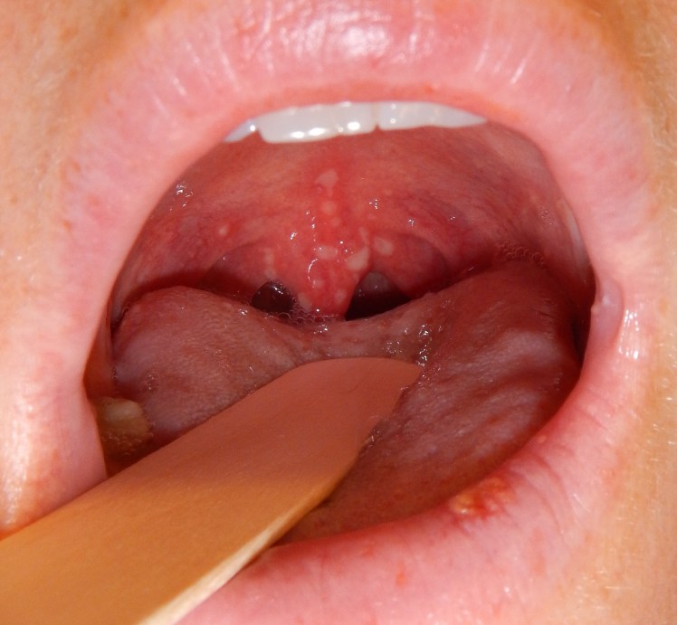
Treatment is supportive. Magic mouthwash (diphenhydramine/Maalox/viscous lidocaine) or ibuprofen for oral pain. Push oral fluids. Ondansetron if vomiting limits intake. Rarely requires admission unless dehydrated.
Dangerous Rashes
Meningococcemia: Rapidly progressive petechiae and purpura in a febrile, toxic child. Can evolve from well-appearing to shock in hours. Any febrile child with petechiae and toxic appearance gets empiric ceftriaxone immediately — do not wait for labs.[1]
Stevens-Johnson Syndrome / TEN: Mucocutaneous reaction with targetoid lesions, skin pain, and mucosal erosions (oral, ocular, genital). BSA involvement distinguishes SJS (<10%) from TEN (>30%). Common pediatric triggers: sulfonamides, anticonvulsants (carbamazepine, lamotrigine), NSAIDs. Stop offending agent immediately. Burns-center transfer for TEN.
Toxic Shock Syndrome: Diffuse macular erythroderma (“sunburn rash”) with fever, hypotension, and multiorgan involvement. Staphylococcal or streptococcal. Desquamation of palms and soles 1-2 weeks later. Aggressive fluid resuscitation, source control, and anti-toxin antibiotics (clindamycin).
Scarlet Fever: Group A strep pharyngitis with erythrogenic toxin — “sandpaper” rash with Pastia lines in skin folds, strawberry tongue. Treat with amoxicillin or penicillin for 10 days. Benign with antibiotics but untreated can cause rheumatic fever.
Drug Reactions
Simple drug eruption (morbilliform): Diffuse maculopapular rash 7-14 days after starting a new medication. Pruritic, symmetric, spares mucous membranes. Self-limited after drug discontinuation. The classic “amoxicillin rash” in a child with EBV is NOT a true penicillin allergy.[3]
Red flags distinguishing dangerous from benign drug reactions: mucosal involvement (SJS/TEN), facial edema or blistering (DRESS), high fever with eosinophilia and organ dysfunction (DRESS — onset 2-8 weeks after drug start), skin pain or Nikolsky sign (TEN), and targetoid or dusky lesions.
DRESS syndrome: Facial edema, diffuse rash, fever, eosinophilia, lymphadenopathy, and organ involvement (hepatitis most common). Onset 2-8 weeks after drug initiation. Anticonvulsants and allopurinol are classic triggers. Requires admission and systemic steroids.
Disposition
Admit: Kawasaki disease (for IVIG and echo). SJS/TEN (burn center for TEN). Meningococcemia or sepsis. ITP with active mucosal bleeding or platelets <20k. Toxic shock syndrome. Any toxic child with petechiae/purpura.
Discharge: Benign viral exanthems. HFMD tolerating oral intake. Urticaria without anaphylaxis features. Simple drug eruption after offending agent discontinued. Mechanical petechiae (above nipple line, well child, normal CBC). Return for new petechiae, mucosal involvement, persistent fever >5 days, worsening rash, or toxic appearance.
References
- Paller AS, Mancini AJ. Hurwitz Clinical Pediatric Dermatology. 6th ed. Elsevier; 2022. Elsevier
- McCrindle BW, Rowley AH, Newburger JW, et al. Diagnosis, treatment, and long-term management of Kawasaki disease: a scientific statement from the AHA. Circulation. 2017;135(17):e927-e999. PubMed
- Lott JP, Liu K, Gaddis KJ, et al. Pediatric rash evaluation and management. Pediatr Emerg Care. 2021;37(3):163-170. PubMed
 tydotphrase.com