MDM Templates
Bloody Stool — Benign / Milk Protein Allergy
Infant presents with blood-streaked stool. Well appearing, hemodynamically stable, soft non-tender abdomen without distension or peritoneal signs.
History and exam not consistent with intussusception, volvulus, NEC, Meckel diverticulum, HUS, or surgical abdomen. In a well-appearing infant with blood-streaked stools and otherwise normal exam, milk protein allergy is the most likely diagnosis.[1]
Plan: Dietary modification education — maternal dairy avoidance if breastfeeding, switch to extensively hydrolyzed or amino acid-based formula if formula-fed. Discharge with PCP follow-up within 48 hours. Return precautions for increasing bloody stools, abdominal distension, fever, lethargy, or poor feeding.
Bloody Stool — Concern for Intussusception
Child presents with episodic abdominal pain and bloody stool. Age and presentation concerning for intussusception given episodic symptoms and clinical findings.[2]
Presentation not consistent with simple gastroenteritis, milk protein allergy, or other benign etiology given the episodic nature of pain and associated bloody stool.
If hemodynamically stable: Ultrasound for diagnosis. Pediatric surgery consulted. Plan for air enema reduction. Admit for post-reduction observation.
If peritoneal signs or hemodynamic instability: Emergent surgical consultation for operative management. IV access, resuscitation initiated, NPO.
Bloody Stool — Surgical / Life-Threatening
Ill-appearing child with bloody stool, hemodynamic instability, and concerning abdominal exam. Presentation raises concern for volvulus, NEC, or significant GI hemorrhage requiring emergent intervention.
Plan: Emergent surgical consultation. IV resuscitation with crystalloid and consideration for blood products. Broad-spectrum antibiotics. NPO. Operative management anticipated.
Clinical Education
Age-Based Differential Diagnosis
The differential for bloody stool in pediatrics is age-dependent. The most important question is how old the child is — this narrows the differential significantly.[1]
| Age | Common | Don’t Miss |
| Neonate (0-1 mo) | Milk protein allergy, swallowed maternal blood, anal fissure, NEC (preterm) | Volvulus, Hirschsprung enterocolitis |
| Infant (1 mo-2 yr) | Milk protein allergy, anal fissure, intussusception | Meckel diverticulum, volvulus |
| Child (2-12 yr) | Infectious colitis, Meckel, juvenile polyps, IBD | Intussusception (pathologic lead point), HUS |
| Adolescent | Infectious colitis, IBD, hemorrhoids | HUS, Meckel |
Red Flags — When to Worry
Bilious emesis is volvulus until proven otherwise. This is a surgical emergency — do not wait for imaging if the child is unstable.
Other red flags requiring urgent evaluation: hemodynamic instability or pallor/lethargy, peritoneal signs or rigid abdomen, currant jelly stool with episodic pain (intussusception), prematurity with feeding intolerance and distension (NEC), and bloody diarrhea with renal failure and thrombocytopenia (HUS triad).[1]
The intussusception “triad” of episodic pain, currant jelly stools, and sausage-shaped mass is rarely complete — only about 20% of cases present with all three. A high index of suspicion in the 6-36 month age group is more important than waiting for the classic presentation.[2]
Not Blood — Mimics
Urate crystals (brick dust): Pink or brick-red crystalline deposits on the diaper in neonates. Completely normal — concentrated uric acid from the first few days of life. No clots, no streaking, just a pink stain. Reassure families.
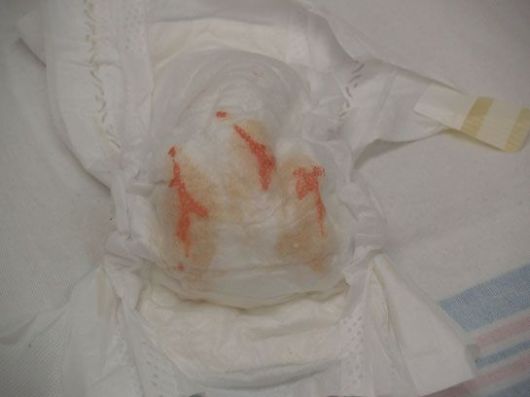
Other mimics: Medications (iron causes black stool, bismuth causes dark stool), red foods (beets, red Jello, Kool-Aid), vaginal bleeding in newborns (normal maternal hormone withdrawal — resolves in 1-2 weeks), and swallowed maternal blood from delivery or cracked nipples during breastfeeding. The Apt test distinguishes fetal from maternal hemoglobin if swallowed maternal blood is suspected.[3]
Intussusception Pearls
Peak age is 6-36 months. In children >3 years, suspect a pathologic lead point (lymphoma, Meckel, polyp) rather than idiopathic intussusception. Ileocolic type accounts for >95% of cases.[2]
Ultrasound is the imaging of choice — sensitivity and specificity >98%. Target sign on transverse view, pseudokidney sign on longitudinal. Do not delay ultrasound for plain films.
Air enema is both diagnostic and therapeutic with approximately 80% successful reduction rate. Post-reduction observation for 24 hours is standard — recurrence rate is approximately 10%, with most recurrences within the first 24 hours.[2]
Surgical indications: perforation, peritonitis, hemodynamic instability, failed enema after 2-3 attempts, or confirmed pathologic lead point.
Milk Protein Allergy
Most common cause of bloody stool in well-appearing infants <6 months. Non-IgE mediated cell-mediated immune response to bovine milk proteins. Typically presents as blood-streaked stools in an otherwise thriving infant.[3]
Management: Breastfed infants — maternal dairy avoidance. Formula-fed infants — switch to extensively hydrolyzed formula (Nutramigen, Alimentum) or amino acid-based formula (Neocate, EleCare). Symptoms typically resolve within 1-2 weeks of dietary change. Most infants tolerate reintroduction by age 1 year.
Refer to GI if: failure to thrive, severe or persistent symptoms despite dietary changes, or no improvement within 3-4 weeks.
Meckel Diverticulum
Most common cause of significant painless lower GI bleeding in children. The rule of 2s: 2% of population, 2 feet from ileocecal valve, 2 inches long, presents before age 2. About 50% contain ectopic gastric mucosa that causes peptic ulceration of adjacent ileal mucosa.[4]
Diagnosis: Meckel scan (technetium-99m pertechnetate scintigraphy) — sensitivity approximately 85% in children (detects ectopic gastric mucosa). Not detected by standard endoscopy since the diverticulum is in the mid-small bowel.
Treatment is surgical resection of the diverticulum and adjacent ileum.
References
- Boyle JT. Gastrointestinal bleeding in infants and children. Pediatr Rev. 2008;29(2):39-52. PubMed
- Waseem M, Rosenberg HK. Intussusception. Pediatr Emerg Care. 2008;24(11):793-800. PubMed
- Koletzko S, Niggemann B, Arato A, et al. Diagnostic approach and management of cow’s-milk protein allergy in infants and children: ESPGHAN GI Committee practical guidelines. J Pediatr Gastroenterol Nutr. 2012;55(2):221-229. PubMed
- Sagar J, Kumar V, Shah DK. Meckel’s diverticulum: a systematic review. J R Soc Med. 2006;99(10):501-505. PubMed
 tydotphrase.com